
Overview

Our dehydrated amniotic membrane allografts are flat and available in disc, rectangle or square shapes. The origins of our product begin with an act of a placental donation from a birth mother giving through a planned Cesarean delivery. The Amniotic Membrane itself is avascular and contains cytokines and growth factors (epidermal, keratinocyte, transforming growth factor beta, vascular endothelial growth factor and platelet derived growth factor). It is in these key growth factors that ocular tissue growth and healing are facilitated. Amniotic Membranes also have anti-microbial and anti-inflammatory properties. 

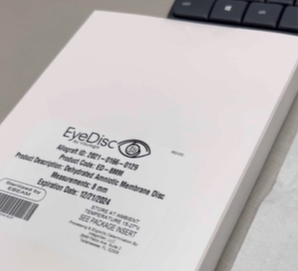
All of our tissues are chorion free and screened for diseases before treating them with broad-spectrum antibiotics.  We provide our dehydrated amniotic membrane allografts in  products from human amniotic tissue in 5 different disc oriented sizes – 5mm, 8mm, 12mm and 16mm; and 3 different EyeGraft sizes 1.5x2cm, 2x3cm and 4x4cm.

Amnion and Amniotic Tissue

EyeDisc and EyeGraft suite of products are amniotic tissue derived allografts intended for reconstruction, repair or replacement of a donor recipients tissue on the ocular surface. In both surgical and clinical applications, EyeDisc and EyeGraft products provide the ocular surface with a tissue barrier and natural regenerative healing properties while simultaneously providing lubrication.

As a Protective Barrier…
During fetal development, Amniotic tissue acts as an immune privileged protective barrier. When applied to the cornea, EyeDisc and EyeGraft offers that same mechanical protection while also providing a regenerative tissue matrix with specific anti-inflammatory, anti-scarring and anti-microbial properties.

In Natural Regenerative Healing…
Because of the notable absence of fibroplast and inflammatory cell activity in the fetal wound healing environment, regenerative healing takes place versus scar mediated healing. EyeDisc and EyeGraft are processed to preserve those exact natural regenerative healing properties. On the ocular surface, endogenous growth factors present in amniotic tissue promote healthy ocular epithelial tissue formation while inhibiting fibrogenesis.

Per Safety and Versatility…
Amniotic tissue is recovered from young, healthy mothers who have undergone cesarean delivery, and is processed in accordance with FDA regulations and AATB standards. Amniotic tissue has been used for over 100 years with excellent clinical success.

Patient Application of EyeDisc

Applying the EyeDisc to the eye is fairly simple and consists of two basic methods outlined below. Important note….the EyeDisc is orientation neutral in its application, meaning that either side of the membrance can be placed onto the corneal surface. EyeDisc will be publishing it’s own set of videos and infographics for patient application in the near future. For now, there steps should be easy to follow.

Application Option A (For the Pragmatist)

  • Place topical proparacaine on the ocular surface
  • Open a bandage contact, take it out and place on sterile space
  • Open the amniotic membrane pouch slowly until the amniotic membrane lens becomes exposed
  • Using forceps, remove the lens from the pouch and place inside of the bandage contact lens
  • Smooth the amniotic membrane disc into the contact lens
  • Prepare the ocular surface of the patient as you normally would prior to a contact lens insertion
  • Place the bandage contact lens onto the corneal surface
  • Complete the enclosed Tissue Tracking Form and return it to our Tissue Bank

Application Option B (For The Purist):

  • Recline patient and tilt chin up, then place topical proparacaine on the ocular surface
  • Insert speculum to hold open eye and dry cornea
  • Open the amniotic membrane pouch slowly until the amniotic membrane lens becomes exposed
  • Using forceps, remove the amniotic membrane lens from the pouch and place over cornea
  • Using forceps still, make sure the amniotic membrane lens is smoothly covering cornea and / or flat to cornea
  • Place bandage contact onto the corneal surface
  • Remove speculum
  • Complete the enclosed Tissue Tracking Form and return it to our Tissue Bank

Materials Needed

  • Blunt Tip Sterile Forceps
  • Proparacaine (or bio-similar topical anesthetic drop)
  • Weck-Cel or Soft-Cel Spear Q-Tips
  • Sterile gloves
  • Sterile work space
  • Bandage Contact Lens